A normal chest x-ray in dogs is a diagnostic modality used by veterinarians to visualize the tissues, organs, and bones within the chest cavity. It is an important tool in identifying any abnormalities or diseases. In a normal chest x-ray, the heart, lungs, blood vessels, and bones should appear healthy and in their proper positions. The x-ray can also help rule out certain diseases and assess the overall health of the dog.
Key Takeaways
- A normal chest x-ray in dogs is used to assess the health of the chest cavity.
- A normal x-ray should show a healthy heart, lungs, blood vessels, and bones.
- Chest x-rays can help rule out diseases and assess overall health.
- Veterinarians use chest x-rays to diagnose and monitor canine health.
- Interpreting a normal chest x-ray involves evaluating the size, shape, and position of structures within the chest cavity.
Why are Chest X-Rays Important for Dogs?
Chest x-rays play a critical role in veterinary medicine, particularly when it comes to dogs. As our four-legged companions are unable to communicate their medical problems, these imaging studies provide valuable insights for veterinarians. By examining the chest cavity, x-rays allow for a comprehensive assessment of vital structures such as the lungs, heart, and surrounding tissues. This non-invasive procedure helps identify various abnormalities, including tumors, fluid accumulation, pneumonia, heart enlargement, and fractures.
Dogs experiencing difficulty breathing, suspected heart or lung diseases, or major trauma often undergo chest x-rays to aid in diagnosis and treatment planning. These images can reveal crucial information about the dog’s underlying health and provide veterinarians with important data to formulate effective treatment strategies. Additionally, regular chest x-rays may be recommended as part of routine diagnostic procedures to ensure ongoing wellness and detect any early signs of disease.
Ultimately, chest x-rays are a powerful tool in the veterinarian’s arsenal, enabling them to have a clearer picture of a dog’s internal health and make informed decisions that can improve their overall well-being.
What Can a Normal Chest X-Ray Reveal?
A normal chest x-ray in dogs can provide valuable insights into their overall health and help identify any potential abnormalities or signs of disease. When interpreting a normal chest x-ray in dogs, veterinarians look for specific findings that indicate healthy anatomy and functioning of the lungs, heart, blood vessels, and bones.
The following are some of the key findings that should be observed in a normal chest x-ray:
- A normal-sized heart: The heart should be within normal size limits, indicating proper functioning.
- Clear and healthy lung fields: The lung fields should appear clear without any signs of congestion, masses, or infiltrates.
- Normal blood vessels: The blood vessels should be well-defined and show no signs of enlargement or abnormalities.
- Well-defined bones: The bones of the chest cavity should be clearly visible with no fractures or signs of structural abnormalities.
By carefully evaluating these structures, veterinarians can detect any changes in size, shape, or position that may indicate potential health issues. A normal chest x-ray is particularly useful in detecting conditions such as heart enlargement, fluid accumulation in the lungs or pleural cavity, and fractures.
However, it is important to note that a normal chest x-ray may not detect all internal problems. In some cases, additional diagnostic procedures or imaging techniques may be necessary to provide a complete diagnosis.

How is a Chest X-Ray Done for Dogs?
To perform a chest x-ray on a dog, specialized and expensive equipment is required. The process involves several steps:
- The dog’s chest is measured and marked to ensure accurate positioning.
- The exposure time of the x-ray machine is set based on the dog’s size and the desired level of detail.
- The dog is gently positioned for the x-ray. This typically involves obtaining both lateral and ventrodorsal views of the chest, which allow for a comprehensive study of the internal structures.
- The x-rays pass through the dog’s body and onto an x-ray film or digital detector, creating images of the chest cavity.
- The films or digital images are then developed and evaluated by a veterinarian or radiologist for interpretation.
Sedation or anesthesia is usually not required for a routine chest x-ray unless the dog is uncooperative during positioning. However, in some cases where the dog is anxious or requires immobilization, sedation may be considered to ensure the safety and accuracy of the procedure.
It’s worth noting that the positioning and technique used during the x-ray can greatly influence the quality and diagnostic value of the images. Skilled veterinarians and radiologists pay careful attention to these factors to obtain the best possible outcomes.
Interpreting a Normal Chest X-Ray in Dogs
The interpretation of a normal chest x-ray in dogs involves a careful evaluation of the structures within the chest cavity, including the bones, heart, lungs, and blood vessels. A radiograph that shows a normal chest x-ray should demonstrate healthy anatomy, with no signs of enlargement, fluid accumulation, or fractures.
When interpreting a normal chest x-ray, it is important to consider the position and orientation of the x-rays. The quality and clarity of the images play a crucial role in identifying any abnormalities or signs of disease. Radiographic abbreviations and terminology may be used to describe specific structures or findings, providing precise details for accurate diagnosis and treatment planning.
A comprehensive interpretation of a normal chest x-ray takes into account the entire image and correlates it with the dog’s clinical history and physical examination. By combining these factors, veterinarians can fully understand the dog’s health status and make informed decisions regarding their care.
“Interpreting a normal chest x-ray in dogs requires careful observation and consideration of various factors. By examining the structures within the chest cavity and analyzing the radiographic images, veterinarians can gain valuable insights into the dog’s health and well-being.”
Importance of Detailed Interpretation
While a normal chest x-ray indicates healthy anatomy, it is essential to review the radiographic images thoroughly. This detailed interpretation allows veterinarians to identify any subtle abnormalities that may not be immediately apparent. Such findings can play a crucial role in detecting early signs of disease, guiding further investigation, and providing timely intervention.
By evaluating a normal chest x-ray in dogs, veterinarians can establish a baseline for future comparisons. Any progressive changes in the subsequent x-rays can be identified and monitored over time. This proactive approach ensures the early detection of potential health issues and enables the implementation of appropriate treatment strategies.
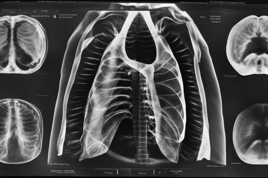
Visualization of Normal Structures
A normal chest x-ray in dogs should reveal the following:
- Healthy bones within the chest cavity, including the ribs and spine.
- A normal-sized heart, indicating no signs of enlargement or abnormality.
- Clear and healthy lung fields, demonstrating proper aeration and absence of fluid accumulation or pathological changes.
- Well-defined blood vessels, indicating no signs of obstruction or abnormalities.
The radiographic images provide insights into these structures, allowing veterinarians to assess the overall health of the dog’s chest cavity.
Conclusion
A normal chest x-ray is an invaluable diagnostic tool for understanding and monitoring the health of dogs. By providing insight into the structures within the chest cavity, it enables veterinarians to detect and evaluate abnormalities that may indicate underlying diseases or conditions. Interpreting a normal chest x-ray involves a comprehensive assessment of the heart, lungs, blood vessels, and bones, considering their size, shape, and position. This requires the expertise and experience of a veterinarian to make informed decisions and provide proper care for the dog.
In routine diagnostic procedures or when there are specific concerns or symptoms related to the chest area, performing a chest x-ray is highly recommended. It allows for a thorough evaluation of the dog’s health, aiding in the early detection and treatment of potential issues. With accurate interpretation and analysis of the x-ray findings, veterinarians can ensure the optimal well-being of their patients.
Therefore, understanding the importance of a normal chest x-ray in dogs and its role in diagnosing and monitoring their health is key. By utilizing this valuable diagnostic modality, veterinarians can provide the best possible care for their canine patients, identifying any abnormalities and developing tailored treatment plans to address specific conditions. Regular chest x-rays, when necessary, contribute to the overall health and longevity of dogs, promoting a happy and active life.
FAQ
What is a normal chest x-ray in dogs?
A normal chest x-ray in dogs is a diagnostic modality used by veterinarians to visualize the tissues, organs, and bones within the chest cavity. It should show healthy anatomy and no signs of abnormalities or disease.
Why are chest x-rays important for dogs?
Chest x-rays are important for dogs as they allow veterinarians to assess the health of the lungs, heart, and other structures within the chest cavity. They can help identify abnormalities such as tumors, pneumonia, and heart enlargement.
What can a normal chest x-ray reveal in dogs?
A normal chest x-ray in dogs can reveal healthy anatomy, including a normal-sized heart, clear and healthy lung fields, normal blood vessels, and well-defined bones. It can also detect changes in the size, shape, or position of these structures, helping to identify abnormalities or signs of disease.
How is a chest x-ray done for dogs?
To perform a chest x-ray on a dog, specialized and expensive equipment is required. The dog’s chest is measured, and the exposure time of the x-ray machine is set. The dog is then gently positioned to obtain both lateral and ventrodorsal views of the chest, which provide a comprehensive study and allow for accurate interpretation.
How is a normal chest x-ray in dogs interpreted?
The interpretation of a normal chest x-ray in dogs involves evaluating the structures within the chest cavity, including the bones, heart, lungs, and blood vessels. The radiographs should demonstrate healthy anatomy, with no signs of enlargement, fluid accumulation, or fractures. The entire image is considered, along with the dog’s clinical history and physical examination.
